
Presentation
Parkinsonism. CT demonstrated an abnormality.
Patient Data


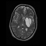
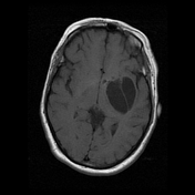
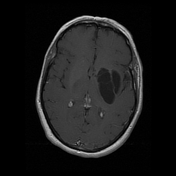
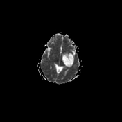
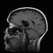
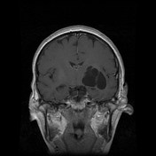
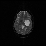
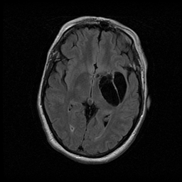
There is a cystic focus measuring 33 x 51 x 35 mm (AP x trans x cc) with apparent septations or opposing cystic spaces centered upon the left basal ganglia, with no evidence of contrast enhancement at this site or elsewhere. There is complete suppression of lesion signal on FLAIR imaging.
There is no significant mass effect, suggesting this lesion is longstanding. There is no surrounding T2/FLAIR hyperintensity to suggest surrounding vasogenic edema.
There is some increased intrinsic T1 signal within the substantia nigra and some bilateral signal loss on susceptibility-weighted imaging within the substantia nigra and red nuclei.
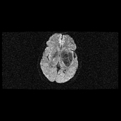
There is no abnormal focus of diffusion restriction with ADC depression.
Elsewhere, no mass or collection is seen. No hydrocephalus or midline shift.
Conclusion:
Cystic left basal ganglia lesion without mass effect, surrounding edema or enhancement, consistent with 'tumefactive' perivascular spaces.
Case Discussion
Tumefactive perivascular spaces can be very striking. The key to the diagnosis is the absence of solid component or surrounding gliosis / edema.


 Unable to process the form. Check for errors and try again.