
Presentation
Abnormality seen on CT, for further assessment.
Patient Data
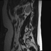

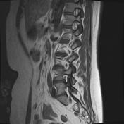
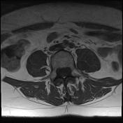
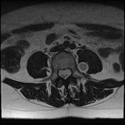

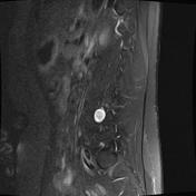
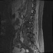
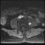
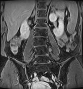

There is a soft tissue mass just lateral to the left L3/4 foramen intimately associated with the L3 nerve root in the lumbar plexus.
The mass demonstrates high signal on T2-weighted sequences, and low signal on T1-weighted sequences. There is a more fluid peripheral component with a solid central component and the lesion measures 2.0 X 1.6 X 1.7 cm.
There is avid contrast enhancement of the central component.
These appearances are consistent with a nerve sheath tumor of the left L3 nerve root within the lumbar plexus in the medial aspect of the psoas muscle. The target appearance on T2 strongly favors a neurofibroma.
Acquired on a 1.5T Toshiba Titan.
Case Discussion
The presence of a target sign is suggestive of a neurofibroma rather than a schwannoma or malignant peripheral nerve sheath tumor, but is unfortunately not pathognomonic.


 Unable to process the form. Check for errors and try again.