
Presentation
An asymptomatic female came for a routine health check-up.
Patient Data
Age: 45 years
Gender: Female
From the case:
Multiple hepatic hemangiomata
Download
Info
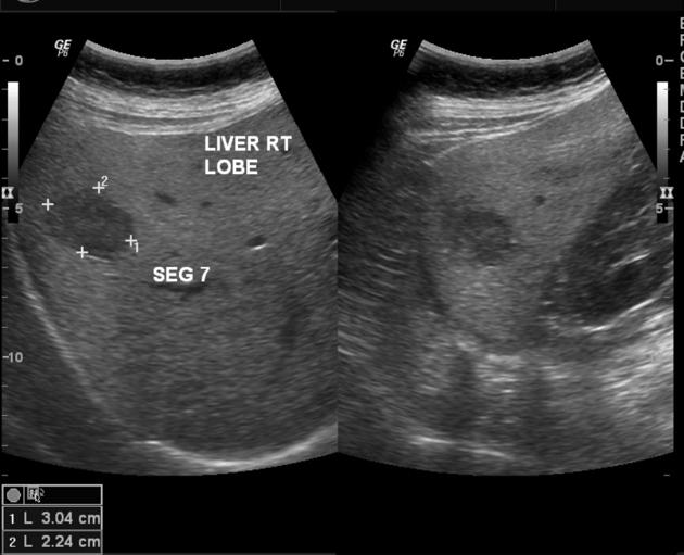
Abdominal ultrasound revealed multiple large heterogeneously hypoechoic mass lesions in both lobes. Largest lesion was over 6 cm in size.
No other positive finding in the abdomen. No abnormality on chest x-ray or mammogram.
A differential diagnosis including hemangiomas, secondary malignancy with occult primary lesion, and primary hepatocellular carcinoma with metastases was given.
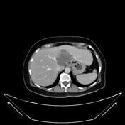
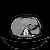
She then underwent a contrast-enhanced CT of the abdomen.
From the case:
Multiple hepatic hemangiomata


Download
Info
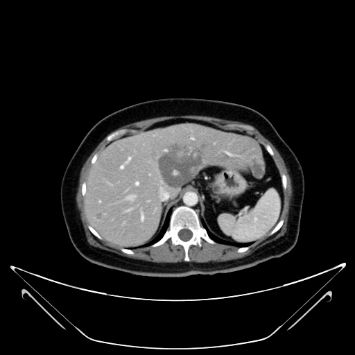
Arterial phase shows peripheral nodular enhancement.
Delayed phase showing complete 'filling in' of the lesions by contrast enhancement.
Case Discussion
A final diagnosis of multiple hepatic hemangiomas was given.


 Unable to process the form. Check for errors and try again.