
Presentation
Young male. Out of hours trauma. Cause unspecified.
Patient Data

Acute right extra-dural hematoma.
Acute left subdural hematoma.
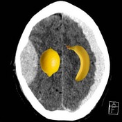


The shape is the key to help differentiate between extradural and subdural hematomas as the fruits illustrate.
Banana image in public domain. Original file: http://imagespng.com/Detail/2011/Banana--PNG-Transparent-image-.html
Lemon image in public domain. Original file: http://imagespng.com/Detail/501/Lemon-PNG-Transparent-image.html
Case Discussion
Following head trauma, CT is usually performed if clinical conditions merit imaging. The major concern is always blood, in particular abnormalities amendable to outcome influencing surgical procedures. This particularly relates to extra-axial collections - extradural and subdural hematomas.
How does one know which is which? The answer is fruity! The shape is usually the key:
- extra-dural hematoma is lentiform like a lemon
- subdural hematoma is sickle shaped like a banana
Image with thanks to Dr I Waldron, Consultant Radiologist, Doncaster Royal Infirmary, UK.


 Unable to process the form. Check for errors and try again.