
Presentation
Fall onto outstretched hand while walking.
Patient Data

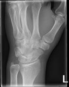

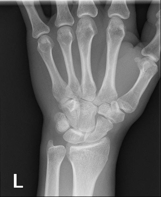
The pronator fat pad is elevated and there is soft tissue swelling over the dorsal aspect of the carpus. There is strong suspicion for a distal radius fracture but one is not identified.


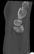
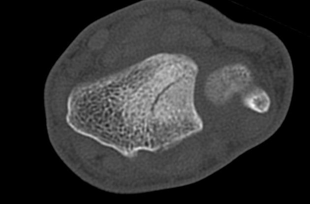
Undisplaced fracture of the distal radius with intra-articular extension. Bulging of the pronator quadratus fat pad. No further fracture is seen.

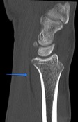
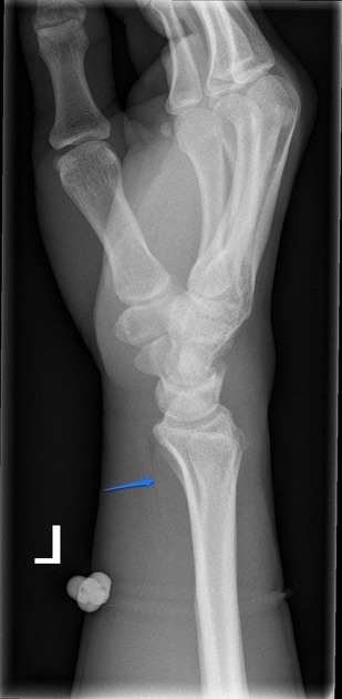
Blue arrows indicate the position of the pronator quadratus fat pad on both plain radiographs and CT. It is bulging outwards (it normally should be straight in plane with the distal radius), which is abnormal.
Case Discussion
This case demonstrate positive pronator quadratus fat pad sign, which can indicate an occult distal fracture (although on its own is non-specific).


 Unable to process the form. Check for errors and try again.