
Presentation
Progressive joint pains.
Patient Data

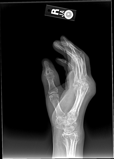
Multiple periarticular erosions are observed bilaterally with adjacent large soft tissue masses and relatively preserved joint spaces.
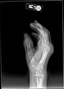
In the right hand, these findings are most prominent at the 1st interphalangeal, 2nd-4th proximal interphalangeal, 1st-3rd metacarpohalangeal, carpometacarpal, intercarpal, radiocarpal and ulnocarpal joints.
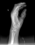
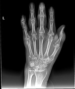
In the left hand, the findings are most prominent at the ulnar styloid, scapholunate joint, first and fifth carpometacarpal joints, second and fifth metacarpophalangeal joints, 1st interphalangeal joint, 3rd, 4th and 5th proximal interphalangeal joints and 3rd and 5th distal interphalangeal joints.

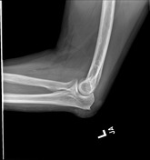
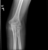
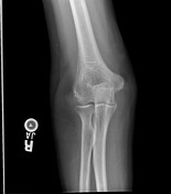
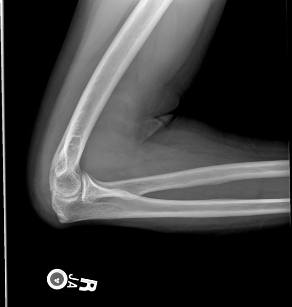
The olecrenon bursae are enlarged bilaterally, with mild erosion of the right olecrenon process. The mass at the left olecrenon bursa has faint areas of calcification.
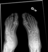
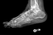
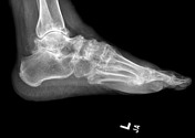

Multiple periarticular erosions are observed bilaterally with adjacent soft tissue masses and relatively preserved joint spaces. The erosions observed throughout the metatarsal bases bilaterally, as well as throughout the midfoot and hindfoot on the left are likely due, in part, to intraosseous tophi.
The distal phalanx of the first digit on the right is dislocated dorsally.
Atherosclerotic vascular changes are noted bilaterally.
Case Discussion
The findings in this case are typical of severe gouty arthropathy.
Case courtesy of Dr. Deborah Forrester


 Unable to process the form. Check for errors and try again.