
Presentation
Visual field defects.
Patient Data


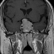

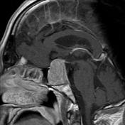
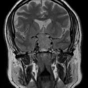
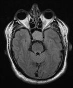
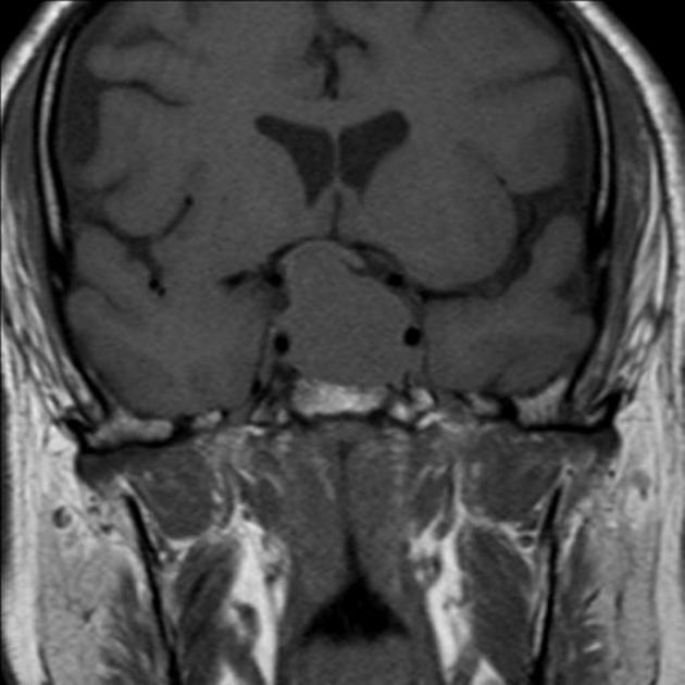
Large homogeneously enhancing mass enlarges the pituitary fossa and compresses the optic chiasm.
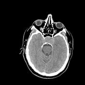
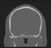
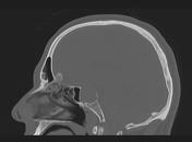

Note is made of an expanded pituitary fossa with a narrowed AP diameter of the pituitary sphenoid sinus. Polyps or mucous retention cysts are present in both maxillary antra with fluid and mucosal thickening in the left ethmoid air cells.
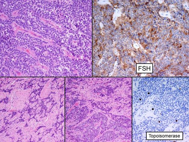
MICROSCOPIC DESCRIPTION: The sections show a moderately cellular pituitary adenoma comprising sheets and nests, surrounded by vascularized stroma. The tumor cells have mildly enlarged round nuclei, finely granular chromatin and moderate amounts of eosinophilic cytoplasm. They infiltrate into the adjacent fibrous tissue, which could be dura. No normal anterior pituitary gland tissue is present. About 75% of the tumor cells are FSH positive. The Ki-67 index is about 3%. Immunostains for the other pituitary hormones are negative.
DIAGNOSIS: Pituitary tumor: "Invasive" silent gonadotroph cell adenoma.


 Unable to process the form. Check for errors and try again.