
Patient Data


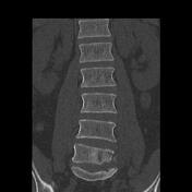
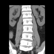
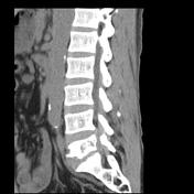

Destructive process involves the left side of the L5 vertebral body. The discs are relatively spared, compared to the degree of bony destruction.
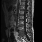
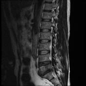
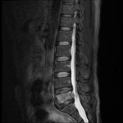
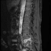
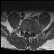
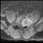
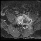
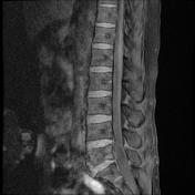

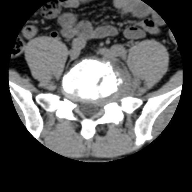
Destructive process involves the left side of the L5 vertebral body, with a non-enhancing fluid component laterally. The discs are relatively spared, compared to the degree of bony destruction. Only a small epidural component is present.
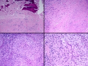


Histology
MICROSCOPIC DESCRIPTION:
The sections show necrotizing granulomatous inflammation with scattered nodular aggregates of epithelioid histiocytes and multinucleated giant cells. Necrosis is seen. The granulomas are surrounded by abundant lymphocytes. No tumor is identified. The Grocott stain shows no fungi. The Ziehl-Neelsen stain shows 2 acid fast bacilli.
DIAGNOSIS:
Spinal lesion: Necrotizing granulomatous inflammation with acid fast bacilli, in keeping with mycobacterial infection.
Microbiology
- ANTIGEN TESTING (ON ISOLATE) MPT64
- Antigen Identification Test: M.tuberculosis Complex DETECTED
- MYCOBACTERIUM CULTURE SCREEN
- MGIT bottle <13 days POSITIVE
- MYCOBACTERIAL CULTURE
- Acid Fast Bacilli ISOLATED


 Unable to process the form. Check for errors and try again.