
Presentation
Chronic knee pain. Medical history of peripheral vascular disease and chronic renal failure.
Patient Data


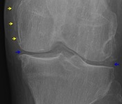
Anteroposterior and lateral radiographs of the left knee demonstrate meniscal calcification (blue arrows) and femoral hyaline cartilage calcification (not labeled). There is also calcification of the medial collateral ligament, quadriceps and patella tendons (yellow arrows). A Pellegrini-Stieda osteophyte is noted in the medial femoral epicondyle.
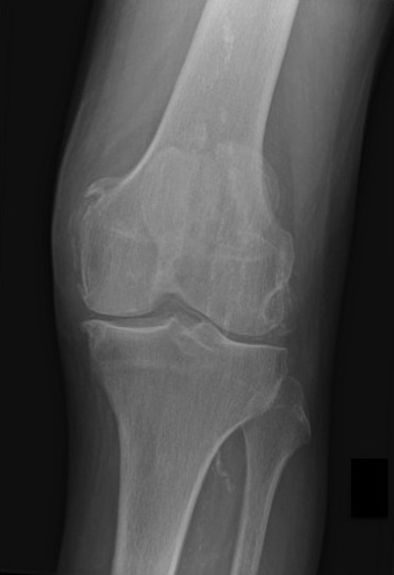
Advanced patellofemoral joint narrowing is accompanied by subchondral bone sclerosis and marginal osteophytes formation.
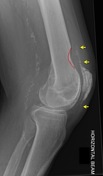
The classic scalloped or "saucerised" remodeling of the ventral metadiaphysis of the distal femur is seen. This is possibly the result of abutment of the patella against the distal femur in knee extension.
Background of extensive vascular calcifications.


 Unable to process the form. Check for errors and try again.