
Presentation
Young patient undergoing work up for newly diagnosed epilepsy.
Patient Data


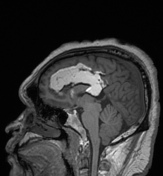
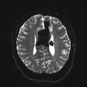
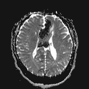
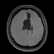

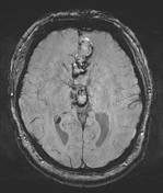
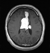

Large lobulated nodular non-enhancing mass in the pericallosal area, with extension into the third and left lateral ventricle. Small amount of SWI susceptibility at the periphery of the mass.
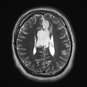
The mass follows fat signal on all sequences with avid fat saturation.
This single set of selected images demonstrates the lesion to be composed of fat.
The fat suppressed T1 sequences shows the extent of the fatty mass.
Case Discussion
Pericallosal lipoma is a fat containing lesion occurring in the interhemispheric fissure closely related to the corpus callosum. It is the most common location for an intracranial lipoma.
Pericallosal lipomas can be divded into two types on the basis of cross sectional imaging studies- tubulonodular or curvilinear.


 Unable to process the form. Check for errors and try again.