
Presentation
Car accident. Thoracic spine pain.
Patient Data


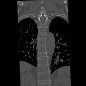

13 paired ribs. The first rib bearing vertebra is taken as T1. Buckling of the superior portion of the T6 and T10 vertebral bodies consistent with acute fractures. Note the angulation of the anterior and lateral cortex at these two levels and subtle compressive sclerosis in the medullary cavity where the normal trabeculae have been impacted. Old vertebral body compression fractures at T9 and L1. Note the remodeled cortex and endplate at these levels without any sharp buckling of the cortex.

White arrows indicate acute vertebral body compression fractures. Yellow arrows indicate chronic vertebral body compression fractures.
Case Discussion
This case demonstrates the differences between acute and chronic vertebral body compression fractures on CT, emphasizing the cortical buckling and subtle trabecular impaction seen with acute fractures compared to the well-defined remodeled appearance of old fractures.


 Unable to process the form. Check for errors and try again.