
Presentation
Knee pain.
Patient Data

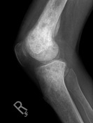
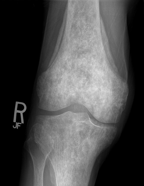
The distal femur and proximal tibia show a diffuse increase in density with a patchy texture. A lamellated periosteal reaction is observed circumferentially around the distal femoral metaphysis, however no focal area of cortical violation or associated soft-tissue mass is observed.
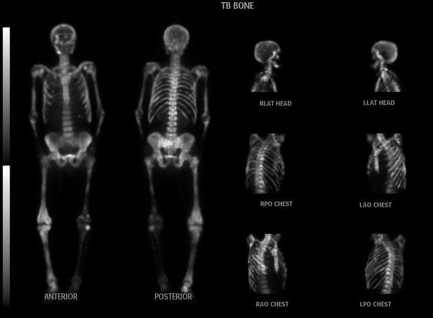
The radio-pharmaceutical uptake in this case is extensive. If it were a little more extensive and more bilaterally symmetric, it might be referred to as a superscan. Notice that almost no radiopharmaceutical is left to be excreted by the kidneys.
Case Discussion
It is a bit unusual to see a periosteal reaction with protate mets, or in any older individual for that matter because the periosteum is tightly adhered to bone and limited in its ability to react quickly or robustly to an underlying pathology. The blastic changes in this case however are none-the-less characteristic, and blastic mets from prostatic carcinoma must be the #1 differential diagnosis in an elderly male with patchy sclerotic bone changes.


 Unable to process the form. Check for errors and try again.