
Presentation
Lower limb symptoms.
Patient Data
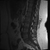

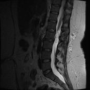
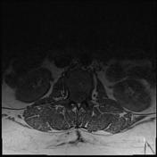
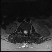

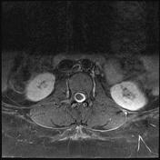

An ovoid intrathecal extramedullary mass is located at the level of the conus, but clearly not arising from it. It has a cystic center and vivid peripheral enhancement.
Case Discussion
It is difficult to be certain of the diagnosis on imaging criteria as both myxopapillary ependymomas and schwannomas can look just like this:
- this other case is an example of a myxopapillary ependymoma
- this other case is an example of a spinal schwannoma
The very vivid enhancement and extremely well defined cystic component probably should make you favor a schwannoma.
The patient went on to have a laminectomy and the mass was excised.
Histology
MICROSCOPIC DESCRIPTION:
Paraffin sections show a partially cystic moderately hypercellular neurilemmoma (Schwannoma). This is composed of both Antoni-A and B type tissues. Moderately well formed Verocay bodies are seen in Antoni-A areas. No mitotic figures are identified. There are no densely hypercellular areas and no areas of necrosis are seen.
DIAGNOSIS: Partially cystic neurilemmoma (Schwannoma).


 Unable to process the form. Check for errors and try again.