
Presentation
Bilateral conductive hearing loss, worse on the right.
Patient Data
Age: 75 years
Gender: Male
From the case:
Fenestral otosclerosis

Download
Info
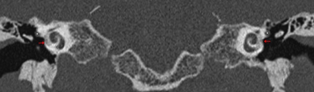
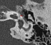
HRCT temporal bone coronal image (A) demonstrates radiolucent halo anterior to the oval window and more pronounced on the right. The tympanic segment of the facial nerve is labeled (*). Axial images (B and C) again shows bilateral radiolucent plaques in the location of the fissula ante fenestram characteristic of fenestral otosclerosis.
Case Discussion
Fenestral otosclerosis is the most common cause of conductive hearing loss in adults and is bilateral in 80% of cases. Early CT changes may subtle as seen in this example on the left side.


 Unable to process the form. Check for errors and try again.