
Nasopharyngeal carcinoma with pterygopalatine fossa involvement
Presentation
Right sided facial pain.
Patient Data


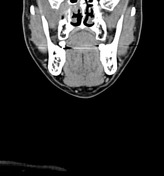
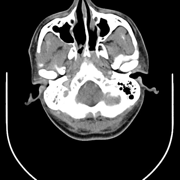
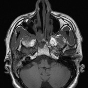
While subtle, there is thickening of the posterior nasopharyngeal wall on the right side. This is associated with opacification of the right mastoid air cells. A salient feature in this case is soft tissue replacement of the normal right pterygopalatine fossa, which is slightly enlarged. Note the normal left side.
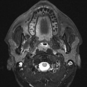

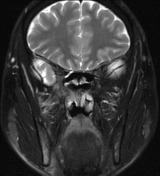
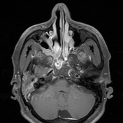

An enhancing soft tissue mass in seen in the right nasopharyngeal cavity. Tumor extends into the right nasopharyngeal cavity, and then intracranially via foramen rotundum and the Vidian canal. There is also extension into the posterior orbit via the inferior orbital fissure.
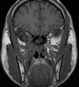
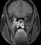
Enlarged bilateral retropharyngeal lymph nodes, and right posterior triangle lymph nodes, are demonstrated.
Case Discussion
The diagnosis of nasopharyngeal carcinoma was proven by pathology.


 Unable to process the form. Check for errors and try again.