Presentation
Inversion injury. Unable to weight bear.
Patient Data
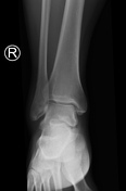
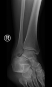
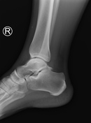
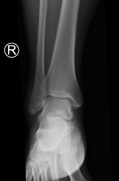
Acute osteochondral fracture at the lateral corner of the talar dome. This is best seen on the AP image, but can be visualized on the lateral and oblique ankle projections too.
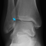

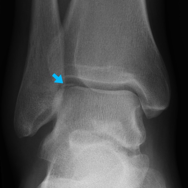
Acute osteochondral fracture at the lateral corner of the talar dome (blue arrow).
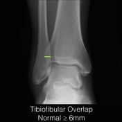

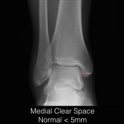
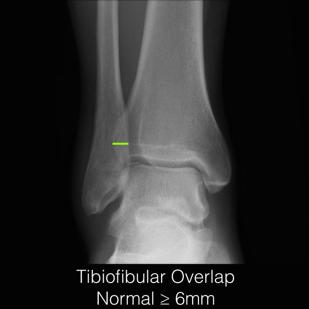
Important normal measurements at the ankle joint:
Tibiofibular overlap should be at least 6 mm on an AP image and usually at least 1 mm on a mortise view. Reduced overlap is a sign of syndesmotic widening/injury.
Tibiofibular clear space should be less than 6 mm on both AP and mortise views. An increase in this space is a sign of syndesmotic widening/injury.
Medial clear space should less than 5 mm on both AP and mortise views. An increase in this space is a sign of deltoid ligament injury (assuming no medial malleolus fracture).
Case Discussion
Acute osteochondral fracture at the lateral corner of the talar dome. This represents an important orthopedic injury as it can often go unnoticed on initial radiography with instability of the osteochondral fragment leading to progressive abnormality and secondary degeneration of the joint.


 Unable to process the form. Check for errors and try again.