
Presentation
Headache and blindness.
Patient Data


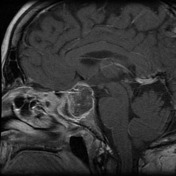
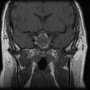
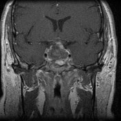
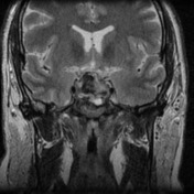
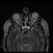
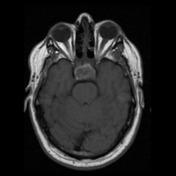
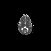
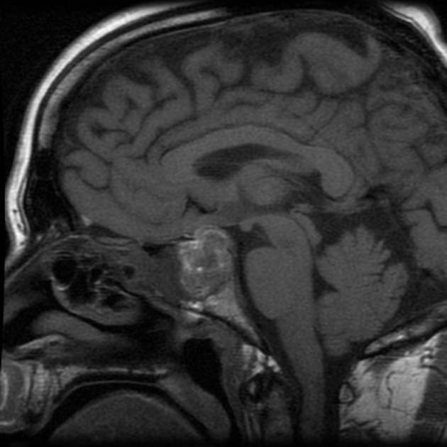
There is a heterogeneous 30 x 23 x 28mm mass centered in the sella extending to the suprasellar cistern. This demonstrates small foci of T1 hyperintensity with central low density on T2 imaging. There is mild peripheral rim enhancement post contrast.
The mass contacts and superiorly displaces the optic chiasm superiorly and displaces the cavernous carotid arteries laterally with mild attenuation of the cavernous carotid flow signal. No phase artifact to suggest this is a pulsatile mass. There is opacification of the sphenoid sinus.
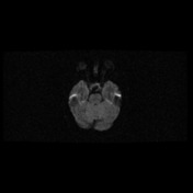
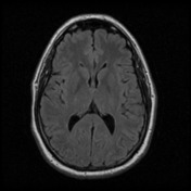
Moderate size area of T2 FLAIR hyperintensity in the right posterior frontal lobe cortex has convex margins, with FLAIR signal extending to cortex. It does not enhance nor diffusion restrict, but note only limited sequences performed as part of this pituitary protocol.
Posterior fossa structures unremarkable. Normal cervicomedullary junction.
MRA (not shown) demonstrates normal opacification of the intracranial ICA, vertebrobasilar system and circle of willis. No aneurysms identified.
Conclusion:
The appearances are more in keeping with pituitary macroadenoma with superimposed hemorrhage/ pituitary apoplexy than a thrombosed large midline aneurysm centered in the sella.
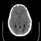


BrainLab Stereotactic CT for Transphenoidal Pituitary surgery
Mildly hyperdense sellar mass seen with suprasellar component. No hydrocephalus.
Nasal septal deviation to left.
Sphenoid sinus septum is Y shaped and roughly 4mm to the right. Sinus roof is deficient posteriorly. Please refer to MRI for ICA course.
Case Discussion
The patient went on to have a transphenoidal resection.
Histology
MICROSCOPIC DESCRIPTION:
Paraffin sections show fragments of extensively necrotic tissue and viable tissue composed of inflamed granulation tissue in which there is prominent reactive fibroblast proliferation. Collections of hemosiderin filled macrophages are also noted. No viable pituitary tissue is identified. The features are consistent with pituitary apoplexy.
DIAGNOSIS:
Pituitary tumor: Necrotic tissue and inflamed granulation tissue consistent with organizing pituitary apoplexy; no viable pituitary tissue identified.


 Unable to process the form. Check for errors and try again.