
Presentation
Hyperparathyroidism
Patient Data


99mTc Sestamibi, 875 MBq
CLINICAL INDICATION: Hyperparathyroidism for localization.
TECHNICAL PROCEDURE AND RESULTS
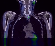
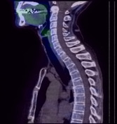
Static and SPECT images were acquired at 15 min and 3 hours. Delayed CT-SPECT was performed.
Initial images show prominent activity at the inferior aspect of the left thyroid lobe. The rest of the thyroid gland shows uniform uptake.
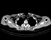
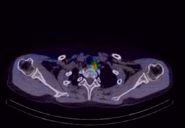
Delayed images show focally retained activity at the inferior aspect of the left thyroid lobe lower pole. SPECT/CT localizes this focal uptake to an intensely Sestamibi-avid soft tissue nodule posteroinferior to the left thyroid lobe, adjacent to T1 vertebral body. The rest of the thyroid gland exhibits normal tracer washout. No other abnormal uptake is detected in the neck or thorax.
OVERALL IMPRESSION
Intensely Sestamibi-avid soft tissue nodule posteroinferior to the left thyroid lobe is consistent with a left lower parathyroid adenoma.
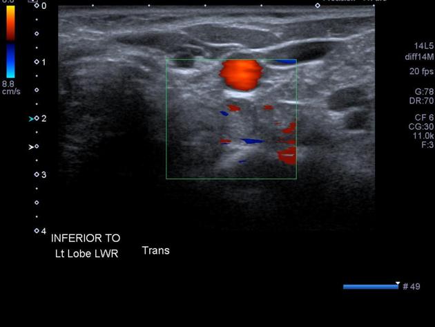
Reference is made to prior examination. Corresponding to the soft tissue nodule posteroinferior to the left thyroid lobe demonstrating intense radiotracer uptake, there is a circumscribed intermediate echogenicity nodule measuring 14 mm, displaying internal color Doppler signal, in keeping with a parathyroid adenoma.
Case Discussion
There is good anatomical correlation between the SPECT and ultrasound localizing the parathyroid adenoma at the lower pole of the left thryoid gland.


 Unable to process the form. Check for errors and try again.