
Presentation
Colorectal cancer. On chemotherapy for liver metastases. Apparent response. Assess for resection
Patient Data

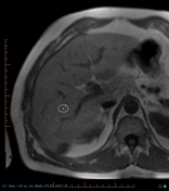
Fatty liver (HU density of 19) showing focal fatty sparing in the posterior left lobe but no mass lesion to indicate metastases.

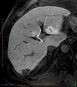


Delayed post Primovist Gadoxetate disodium contrast enhancement contemporary with CT
Marked loss of signal intensity (133 to 47) on the out of phase sequence is due to intravoxel fat (fatty liver). Delayed post Primovist scans of the liver demonstrate multiple non-cystic mass lesions without contrast enhancement but with restricted diffusion indicative of metastases in both lobes.
Case Discussion
As a result of the generalized reduction in parenchymal density of the liver that occurs with fatty infiltration, lower density mass lesions such as metastases can be "lost" as there is very little density difference between the mass and the adjacent fatty liver. Primovist is taken up by hepatocytes in the liver irrespective of fatty change whereas the malignant cells do not, thus there is a signal intensity difference and the lesions are visible. The metastases also show restricted diffusion due to the presence of tightly bound water molecules in the cytoplasm of malignant cells with large nuclei.


 Unable to process the form. Check for errors and try again.