
Patient Data
Age: 55 years
Gender: Male
From the case:
CADASIL (CT only)


Download
Info
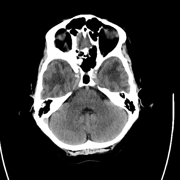
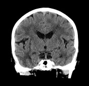
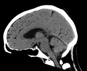
Extensive confluent deep and subcortical cerebral white matter hypo-attenuating changes involving the temporal white matter and the external capsules are noted. Some of established chronic lacunar infarcts demonstrate vacuolation/cavitation. A focal cortical infarct in the lateral aspect of the right postcentral gyrus of undetermined age.
In addition there is ill-defined hypoattenuation change in the left hemi pons likely to reflect ischemic event. Mildly prominent ventricles. No extra-axial or intra-axial acute hemorrhage or collection.
Case Discussion
This patient had a confirmed diagnosis of CADASIL and demonstrates the typical temporal and external capsule dominant chronic small vessel ischemic change.


 Unable to process the form. Check for errors and try again.