Presentation
Swelling to the left side of the neck. Recent hoarse voice. Heavy smoker.
Patient Data
Note: This case has been tagged as "legacy" as it no longer meets image preparation and/or other case publication guidelines.
![](https://prod-images-static.radiopaedia.org/images/11667332/9b5a7a7a10e620bea25c4598c49c69_thumb.jpg)
![](https://prod-images-static.radiopaedia.org/images/11667340/873758698350097767ed2736398a20_thumb.jpg)
![](https://prod-images-static.radiopaedia.org/images/11667341/e17b9dc6adffa8f628886f3cb1ea7d_thumb.jpg)
![](https://prod-images-static.radiopaedia.org/images/11667346/f1b8bd8525706f7488d85eb9b0665c_thumb.jpg)
![](https://prod-images-static.radiopaedia.org/images/11667332/9b5a7a7a10e620bea25c4598c49c69_big_gallery.jpg)
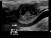
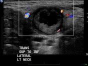
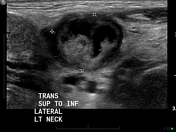
Typical centrally necrotic lymph node on the left side of the neck, confirmed to be metastatic SCC on fine needle aspiration biopsy.
![](https://prod-images-static.radiopaedia.org/images/11667364/4353a0e8b246feab6aa3aefba75885_thumb.jpg)
![](https://prod-images-static.radiopaedia.org/images/11667368/27272b1de9c61248f2dc0d8de0950e_thumb.jpg)
![](https://prod-images-static.radiopaedia.org/images/11667372/1991cc50cf5bf42f9f4c1588fa59f1_thumb.jpg)
![](https://prod-images-static.radiopaedia.org/images/11667381/9519943ee896fba21c159c56ddee5c_thumb.jpg)
![](https://prod-images-static.radiopaedia.org/images/11667387/198f32deb04c7a3331f34b9c5b8a37_thumb.jpg)
![](https://prod-images-static.radiopaedia.org/images/11667364/4353a0e8b246feab6aa3aefba75885_big_gallery.jpg)
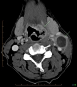
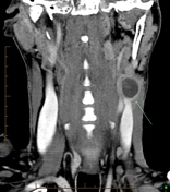
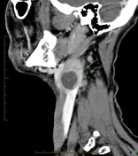
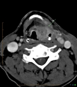
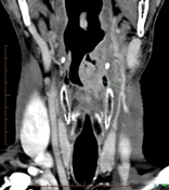
Large malignant appearing mass in the left supraglottic region extending down to the left vocal cord. Palpable node seen on US is confirmed on CT to represent a a rim-enhancing mass consistent with an infiltrated lymph node.
Case Discussion
SCC lymph node metastases typically show central necrosis on ultrasound and CT. If seen in the neck then a head and neck primary malignancy should be suspected. Differential diagnosis is Mycobacterium infection.