
Presentation
Transient right leg weakness and slurred speech. No history of seizures.
Patient Data


No acute intracranial hemorrhage. No CT evidence of evolving infarct. Minor bilateral white matter hypodensities are in keeping with a degree of chronic small vessel ischemia.
No hydrocephalus.
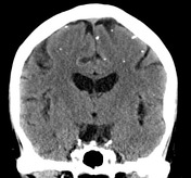
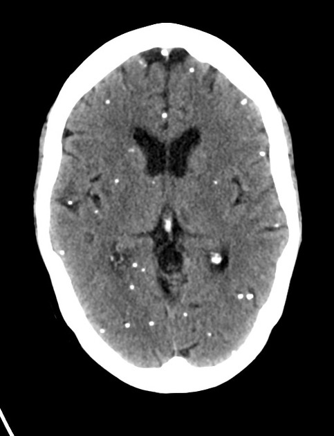
Innumerable subcentimeter calcified lesions lying predominantly throughout the supratentorial brain is in keeping with nodular calcified neurocysticercosis.
Partially empty sella. Prominent retrocerebellar CSF space likely represents either a small arachnoid cyst or mega cisterna magna.
No suspicious bony lesion. Degenerative joint disease noted within the left temporomandibular joint.
Conclusion:
Nodular calcified neurocysticercosis.
No CT evidence of evolving acute cerebral infarct.
Case Discussion
Neurocysticercosis is caused by CNS infection with Taenia solium. There are four main stages of infection: vesicular, colloidal vesicular, granular nodular and nodular calcified. This case represents the end-stage, nodular calcified form of the disease.


 Unable to process the form. Check for errors and try again.