Presentation
Double vision, fevers, and headaches
Patient Data
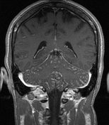


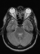



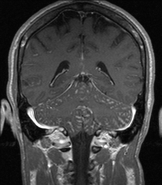
There is marked diffuse leptomeningeal enhancement most pronounced over the cerebellum and in the basal cisterns. There is also nodular enhancement in the right basal ganglia likely via the perivascular spaces.
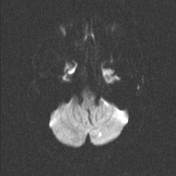
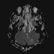
There is a focus of restricted diffusion in the left inferomedial cerebellum compatible with a small PICA branch infarction. There is no hemorrhage.
Small polyps or retention cysts in the maxillary sinuses.
Case Discussion
Etiologies of leptomeningeal enhancement include: infection (viral, bacterial, or fungal), sarcoid, or neoplastic (lymphoma or carcinomatous).
In this case patient had a spinal tap and CSF demonstrated: (reference range)
- Color/appearance: Colorless, clear
- WBC: High 89 (0-10/UL)
- RBC: 12
- Neutrophils: High 11% (0-6%)
- Lymphocytes: High 86% (40-80%)
- Monocytes: Low 3% (15-45%)
- Glucose: 49 (40-70 MG/DL)
- Total Protein: High 118 (15-45 MG/DL)
- Gram Smear: No Cells Seen
- VDRL: nonreactive
- Indian Ink and Culture: Positive for C. neoformans.
The patient was diagnosed with Cryptococcal meningitis. There was no known history of HIV. The patient was also found to have a small left PICA infarct likely secondary to vacuities/spasm.


 Unable to process the form. Check for errors and try again.