
Presentation
Evaluate for stones
Patient Data
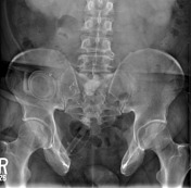

Calcific density anterior to the S1 vertebral body is a stone in the ileal conduit.
Multiple surgical changes secondary to repair of bladder exstrophy are seen in the right lower quadrant.
Widened pubic symphysis, congenital here since there is surgical history it is from exstrophy.
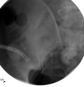
Surgical clips are visualized in the right lower quadrant with colostomy overlying the right iliac crest.
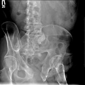
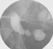
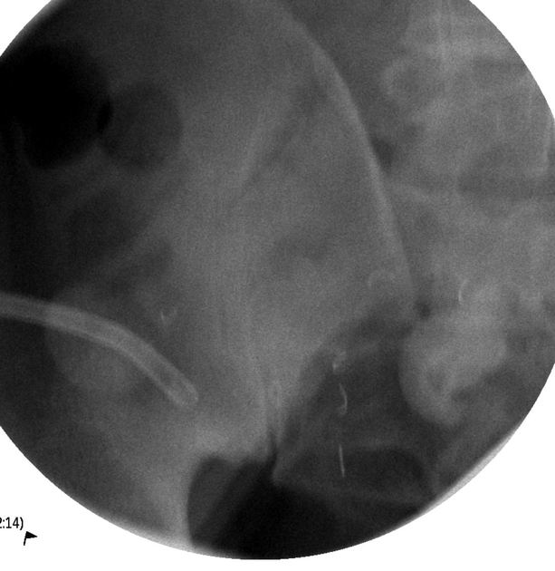
Contrast is injected into the ileal conduit - two irregular-shaped opacities with increased density are seen in the pelvis representing stones in the ileal conduit.
Ostomy is seen.
Case Discussion
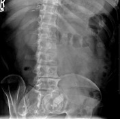
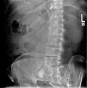
Ileal conduit and ileal conduit stones.
Left nephrostomy placement was requested for access for the stone removal procedure.
There is an increased risk for stone formation in ileal conduits for a variety of reasons.


 Unable to process the form. Check for errors and try again.