From the case:
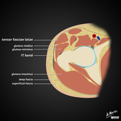
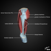
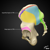
Iliotibial band (ITB) anatomy: diagrams

Download
Info

Diagrams of various components of iliotibial (IT) band anatomy

At the time the case was submitted for publication Matt Skalski had no recorded disclosures.
View Matt Skalski's current disclosuresDiagrams of various components of iliotibial (IT) band anatomy
You can use Radiopaedia cases in a variety of ways to help you learn and teach.
Creating your own cases is easy.
ADVERTISEMENT: Supporters see fewer/no ads
Updating… Please wait.

 Unable to process the form. Check for errors and try again.
Thank you for updating your details.