
Presentation
Follow-up for demyelination disease.
Patient Data


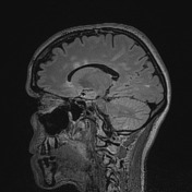


Multiple foci of high T2/FLAIR signal have been again identified scattered within the corpus callosum, at the callososeptal interface, periventricular and subcortical white matter, as well as in the pons and left cerebellar hemisphere.
These have remained stable in appearance and number, with no definite new lesions identified, and no restricted diffusion.
The remainder of the brain is also within normal limits considering the present MRI protocol, with no intra or extraaxial collection, mass or region of abnormal contrast enhancement.
Conclusion:
Patient with a known diagnosis of multiple sclerosis showing stability on MRI findings, with no evidence of current demyelinating activity.
Case Discussion
Multiple sclerosis is a relatively common acquired chronic relapsing demyelinating disease involving the central nervous system. It is by definition disseminated not only in space (i.e multiple lesions), but also in time (i.e. lesions are of different age).


 Unable to process the form. Check for errors and try again.