
Presentation
Left breast painless lump increasing in size over 9 months.
Patient Data
Age: 30 years
Gender: Female
From the case:
Giant fibroadenoma

Download
Info
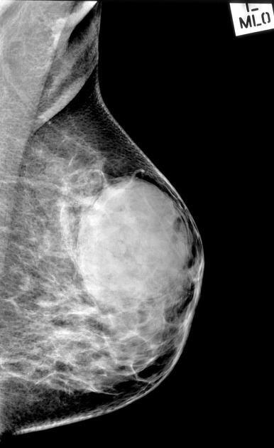
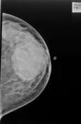
Very large dense mass with well-defined margins, posterior and slightly superior to the nipple.
From the case:
Giant fibroadenoma
Download
Info
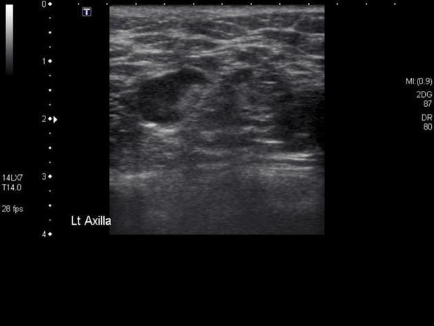
4.5 x 2 .6 cm well circumscribed well defined ovoid mass at the 12 o'clock position 2 cm from the nipple. The mass is predominantly solid, contains multiple small cystic spaces and large areas of echogenic material which appeared to stream (consistent with fluid).
Core biopsy performed.
Case Discussion
The imaging appearance favors a giant fibroadenoma or less likely a phyllodes tumor with necrosis.
Histopathology results were that of fibroadenoma.


 Unable to process the form. Check for errors and try again.