
Presentation
School girl. Active in sport. Twisting injury. Pain and intermittent locking.
Patient Data
Age: 15 years
Gender: Female
From the case:
Osteochondral fracture


Show annotations
Download
Info
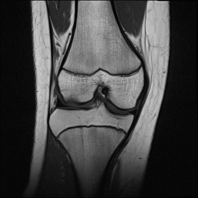
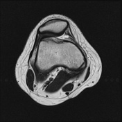
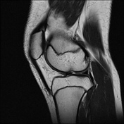
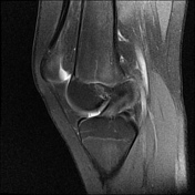
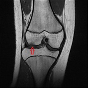
Osteochondral defect on the lateral femoral condyle.
A 7 mm bone fragment lies anteriorly in the lateral joint space.
The remainder of the knee is normal.
From the case:
Osteochondral fracture
Download
Info

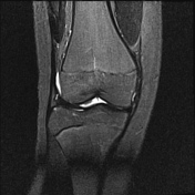
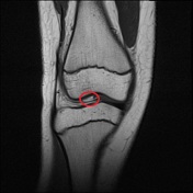
The intra-articular loose body (circle) corresponds with the osteochondral defect (arrow) on the lateral femoral condyle.
Case Discussion
Osteochondral defects are focal areas of articular damage with cartilage damage and injury of the adjacent subchondral bone.
MRI is the modality of choice, with high sensitivity and specificity for the detection of separation of the osteochondral fragment
Osteochondral injuries are staged from I - V. The displaced osteochondral fragment makes this a stage IV injury.


 Unable to process the form. Check for errors and try again.