
Presentation
Acute heart failure.
Patient Data
Age: 30 years
Gender: Female
From the case:
Non-compaction of the left ventricle


Download
Info
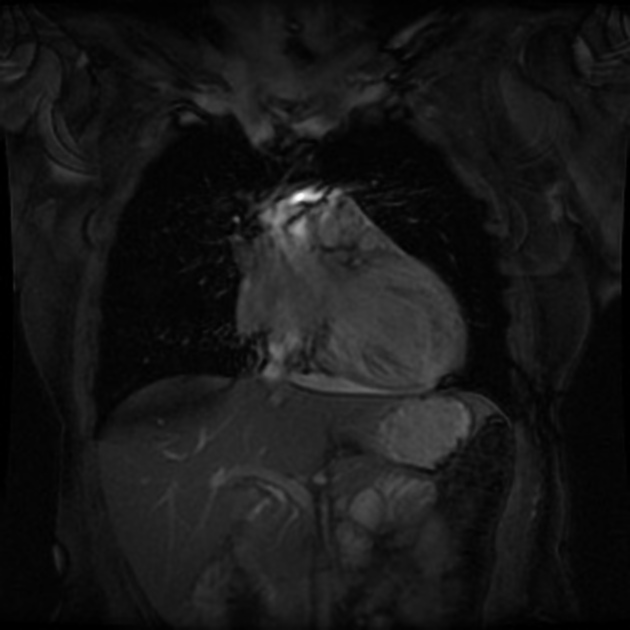
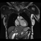
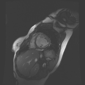
Extensive trabeculations within the left ventricle and thinning of the underlying compact myocardium.
Steady-state free precession cine images in diastole. Abnormal (> 2.3:1) of non compacted to compacted myocardium in diastole.
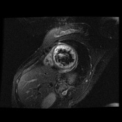
Subendocardial increased signal.
Late gadolinium enhancement image shows subendocardial enhancement consistent with fibrosis at the apex.
From the case:
Non-compaction of the left ventricle
Download
Info
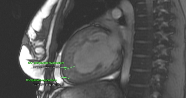
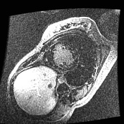
The ratio of the non compaction myocardium to the compacted myocardium is 3 (1.5/.5)
Case Discussion
There is excessive trabeculation at left ventricular myocardium. 2.3:1 ratio of non compacted to compacted myocardium at end-diastole on SAXs CINE or LONG AXIS at the apex.
Differential diagnosis:
- apical hypertrophic cardiomyopathy
- dilated cardiomyopathy
- late gadolinium enhancement imaging with long inversion time (~ 600 milliseconds) to exclude thrombus


 Unable to process the form. Check for errors and try again.