
Presentation
Paroxysmal stabbing facial pain lasting for several minutes.
Patient Data
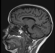

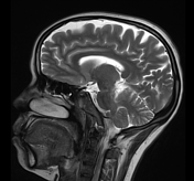
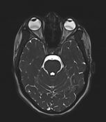

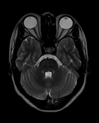
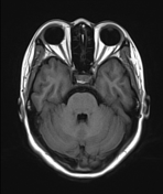
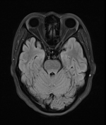
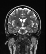

T1, T2, and 3D CISS images show foci of signal void touching the cisternal segment of the right fifth cranial nerve.
3D VIBE image shows tortuous superior cerebellar artery touching the right trigeminal nerve.

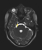
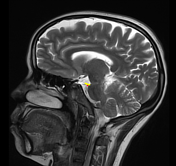
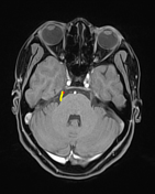
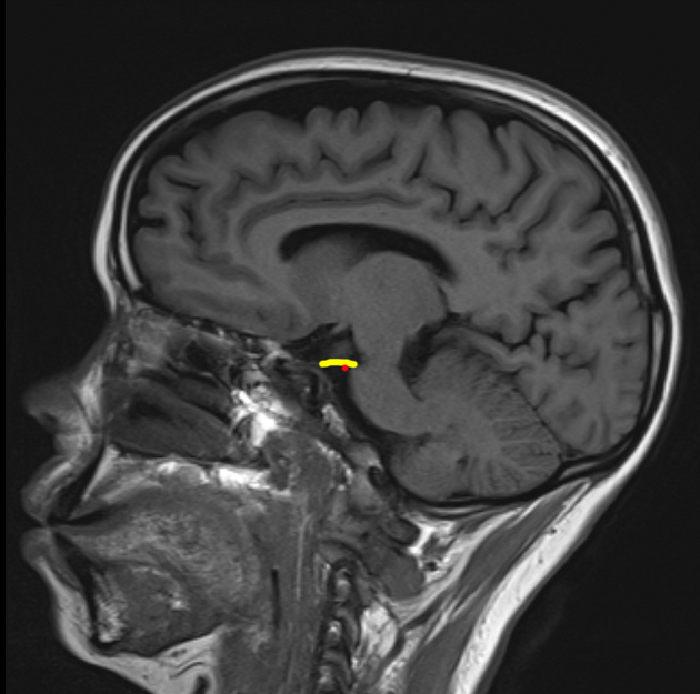
Annotated images T1 sagittal, 3D CISS axial, T2 sagittal, 3D VIBE axial.
yellow - trigeminal nerve.
red - superior cerebellar artery.
Case Discussion
The trigeminal nerve is divided into four segments: brainstem, cistern, Meckel cave/ cavernous sinus and extracranial segments.
The common causes of trigeminal neuralgia (Tic douloureux) are 1:
- brain stem segment: multiple sclerosis, infarct and glioma
- cisternal segment: neurovascular compression, acoustic and trigeminal schwannomas, meningiomas, epidermoid cysts, lipomas and metastases
- Meckel cave and cavernous sinus: meningiomas, trigeminal schwannomas, epidermoid cysts, metastases, pituitary adenoma and aneurysm.
- extracranial: perineural tumor spread
Tortuous, elongated superior cerebellar artery (common), elongated anterior inferior cerebellar artery, vertebrobasilar dolichoectasia or venous compression are the causes of neurovascular compression in the cisternal segment of trigeminal nerve 1.


 Unable to process the form. Check for errors and try again.