
Presentation
Left trigeminal neuralgia.
Patient Data


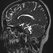
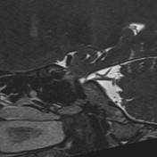
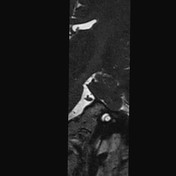

These heavy weighted T2 high-resolution scans of the posterior fossa clearly showing the left superior cerebellar artery indenting the left trigeminal nerve at its inferior surface. No significant discrepancy in girth is seen between the trigeminal nerves on either side.
The left superior cerebellar artery can be traced to its origin from the basilar artery (best demonstrated at the coronal scan). It is seen looping downward and touching the inferior aspect of the trigeminal nerve at its middle segment.
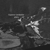


The left trigeminal nerve and superior cerebellar artery are annotated and their relation is clearly demonstrated.
Case Discussion
Neurovascular compression of the trigeminal nerve at the CP angle by the superior cerebellar artery is one of the common causes of trigeminal neuralgia. Classically, in cases of vascular looping, the affected nerve is seen partially encased by the artery, however in this case the artery is clearly abutting the superior surface of the nerve.
The presence of relative nerve thickening (discrepancy in girth) or signal alteration is another confirmatory sign to diagnose a neurovascular compression syndrome.
Although in this case none of these findings is present, the presence of vascular indentation of the nerve supported by a clinical presentation corresponding to the same affected side, in the absence of other pathologies affecting the trigeminal nerve (such as trigeminal schwannoma or any other CP angle mass compressing the nerve), the possibility of the trigeminal neuralgia due to vascular compression must be highly suggested.


 Unable to process the form. Check for errors and try again.