
Presentation
Long-standing history of seizures.
Note: This case has been tagged as "legacy" as it no longer meets image preparation and/or other case publication guidelines.
From the case:
Mesial temporal sclerosis

Download
Info
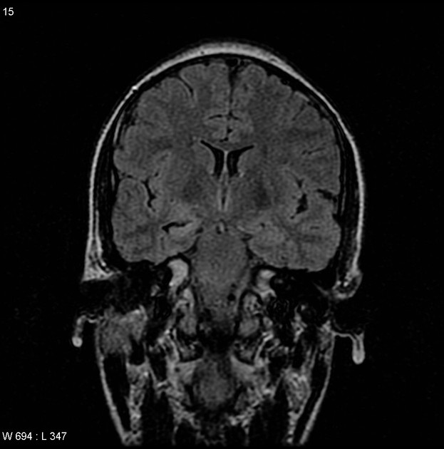
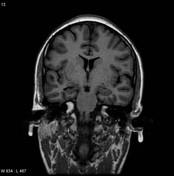
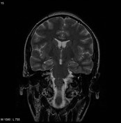
Selected images demonstrate a small right hippocampus with increased T2 signal consistent with right-sided mesial temporal sclerosis.
Case Discussion
The patient went on to have a hippocampectomy.
Histology
Sections of the mesial structures confirm mesial temporal sclerosis, which is represented by a profound depletion of neurons within CA1. A localized aggregate of neurocytic cells is observed, equivalent to focal microdysgenesis. No other diagnostic neuropathologic findings are seen. Subpial gliosis is evident.
Final diagnosis: mesial temporal sclerosis


 Unable to process the form. Check for errors and try again.