
Presentation
Immunosuppressed (post bone marrow transplant). Bilateral sensorineural hearing loss (SNHL).
Patient Data
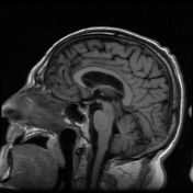

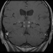
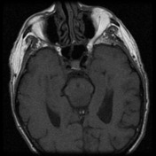

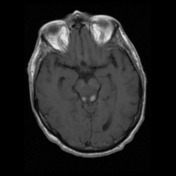
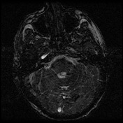
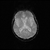
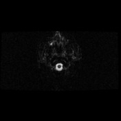
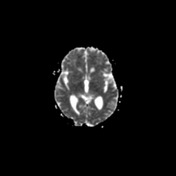

Abnormal FLAIR signal abnormality and expansion within the posterior thalami / pulvinar bilaterally, extending into the posterior midbrain and tectal plates. Discrete ovoid foci of enhancement seen in the superior and inferior colliculi bilaterally as well as within the thalami. The lesions within the inferior colliculi have peripheral rim enhancement. No diffusion restriction.
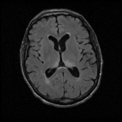
Multiple FLAIR hyperintense foci scattered throughout the white matter are non-specific (unlikely to be related to thalamic/brainstem abnormality) - may represent the sequelae of chronic small vessel ischemic change.
No acoustic neuroma.
Mild paranasal sinus mucosal disease.
Conclusion: Signal abnormality within the thalami and posterior midbrain with discrete foci of contrast enhancement as described, accounting for symptoms of sensorineural deafness. Differential includes encephalitis (both infective and inflammatory - viral and atypical organisms such as CMV, listeria, toxoplasmosis should be considered) as well as neoplastic processes such as lymphoma (less likely glioma).
Case Discussion
Signal abnormality within the thalami and posterior midbrain with discrete foci of contrast enhancement as described, accounting for symptoms of sensorineural deafness. Differential includes encephalitis (both infective and inflammatory - viral and atypical organisms such as CMV, listeria, toxoplasmosis should be considered) as well as neoplastic processes such as lymphoma (less likely glioma).
CSF sampling revealed toxoplasmosis, which is an opportunistic infection caused by parasite Toxoplasma gondii.
Neurotoxoplasmosis has its lesions occurring generally in the basal ganglia, corticomedullary junction, thalamus, and cerebellum.


 Unable to process the form. Check for errors and try again.