
Presentation
Back pain
Patient Data
Age: 25 years
Gender: Female
From the case:
Fused hemivertebra

Download
Info

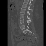
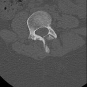
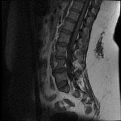
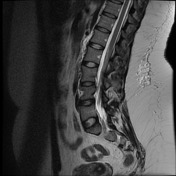
On the right side of L4 and additional hemivertebra is present with the body and lateral mass fused to L4 with resultant dextroscoliosis. The right-sided facet joints demonstrate marked degenerative change.
From the case:
Fused hemivertebra


Download
Info
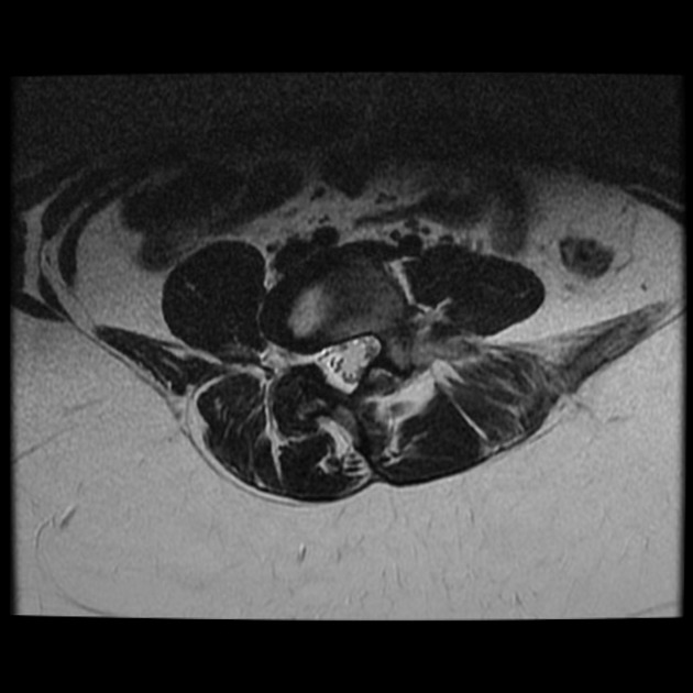
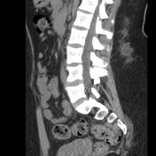
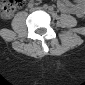
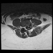
Fused L4 hemivertebra with short segment dextroscoliosis is noted, best seen on coronal images. A nerve root appears to exit via the foramen between L4 and the hemivertebra. No canal stenosis.


 Unable to process the form. Check for errors and try again.