Presentation
Hypertension. Elevated calcitonin.
Patient Data

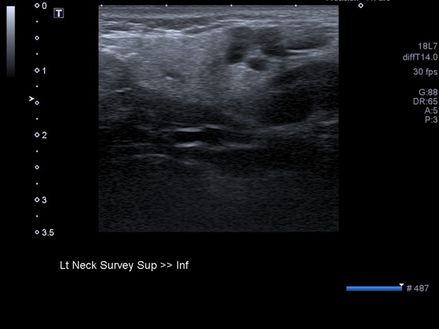
In the midpole the right thyroid lobe is an 11 mm ill-defined hypoechoic lesion with several punctate foci of microcalcification and internal vascularity. In the midpole of the left thyroid lobe is a similar lesion measuring 12 mm.
There are prominent lymph nodes in both sides of the neck, with a 9 mm level IV lymph node on the right which contains punctate foci of microcalcification.
Thyroid FNA
MICROSCOPIC DESCRIPTION: The smears are hypercellular and contain atypical epithelial cells presenting in sheets, and complex crowded tissue fragments. The cells show predominantly spindle-shape morphology with ovoid and elongated nuclei but others have a "plasmacytoid" appearance. The nuclei exhibit stippled chromatin pattern. Nucleoli are inconspicuous. Small globules of metachromatic material are present in the background.
DIAGNOSIS: FNA Thyroid - Left Lobe: Medullary carcinoma.

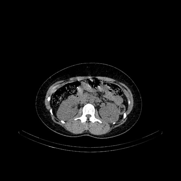
Large bilateral upper abdominal masses of heterogeneous density at the superior aspect of the kidneys. Normal adrenal glands are not seen.


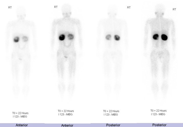
The scintigraphic findings are consistent with bilateral adrenal pheochromocytomas, with markedly increased tracer seen in both adrenal masses.
Patient proceeded to resection of bilateral adrenal masses.
Histopathology:
MACROSCOPIC DESCRIPTION:
1. "Left pheochromocytoma": A 375g solid cystic nodule 90x90x75mm. The tumor has a smooth capsular surface with some attached fat 80x35x15mm. Flecks of orange strands of tissue are present at the edge of the nodule and may represent residual cortex. The tumor itself has a spongy brown cut surface with some cystic areas measuring up to 37mm in maximum dimension present.
2. "Right pheochromocytoma": A 370g nodule 115x85x85mm. The nodule has a smooth capsular surface with some peritoneum covering it focally over a 60x40mm area. The tumor has a similar cut surface to specimen 1 with cystic spaces up to 25mm intermixed with more solid red tan spongy parenchyma. Flecks of orange tissue are present peripherally possibly consistent with residual cortex. Separate to the tumor is a nodule of fatty tissue measuring 15x15x10mm which is loosely attached to the capsule of the tumor.
MICROSCOPIC DESCRIPTION: 1&2: Sections of each of the two specimens show similar features of pheochromocytoma. Tumor cells have pleomorphic round and oval vesicular nuclei and a large amount of granular basophilic cytoplasm and are arranged in diffuse sheets and lobules within an intensely vascular stroma. Scattered mitotic figures are identified (up to 3/20HPF). No necrosis is identified. Tumor appears to be partially delimited by a fibrous capsule in each specimen and is sharply demarcated from the small amount of normal adrenal cortex that is included in specimen 1. No normal adrenal cortex is seen in specimen 2. Extra-adrenal adipose tissue is unremarkable.
DIAGNOSIS: 1&2: "Left & Right pheochromocytoma": Pheochromocytoma x2.
Case Discussion
MEN2 consists of medullary thyroid cancer (always present) and pheochromocytoma (commonly present). It can be further divided into MEN2a with the addition parathyroid hyperplasia and MEN2b with the presence of mucosal neuromas.


 Unable to process the form. Check for errors and try again.