
Presentation
No history provided.
Patient Data


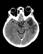
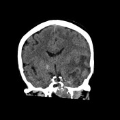
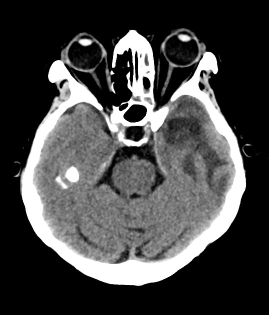
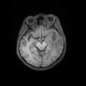
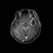
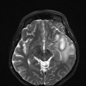
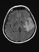
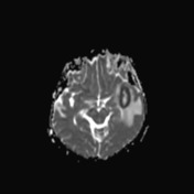
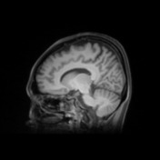
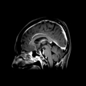
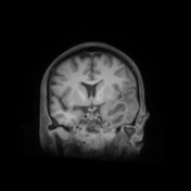
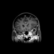
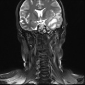
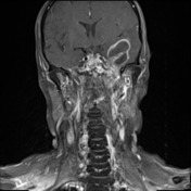
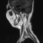
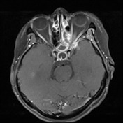
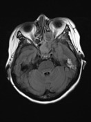
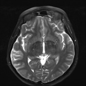
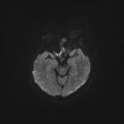
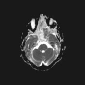
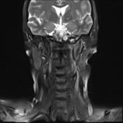
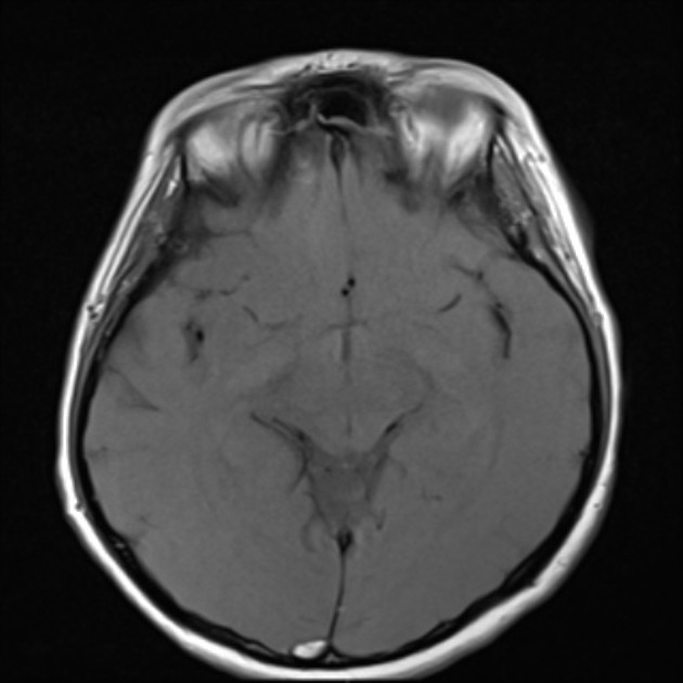
Left temporal lobe rim enhancing, multi-loculated cystic lesion is demonstrated with moderate surrounding white matter hypodensity. Extensively infiltrating lesion is demonstrated in the left skull base, arising from the left side of the nasopharynx and resulting in bony destruction of mainly the sphenoid bone, also just encroaching on the apex of the petrous temporal bone and clivus. The lesion abuts the left carotid canal. The left sphenoid sinus and ethmoid sinuses are partially opacified. The right sphenoid sinus and left maxillary sinus are almost completely opacified. There is moderate mass effect resulting in left frontotemporal sulcal compression and mild compression of the left lateral ventricle. No significant midline shift.
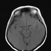
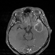
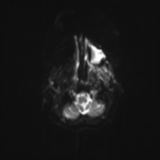

Destructive lesion at the left skull base is compatible with locally invasive nasopharyngeal carcinoma (NPC).
Left temporal lobe multiloculated cystic rim-enhancing, centrally restricting, lesion is compatible with a cerebral abscess. Similar findings are seen in the left maxillary antrum, presumably also infected.

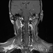

Large destructive left sided nasopharyngeal mass is visible on this scan.
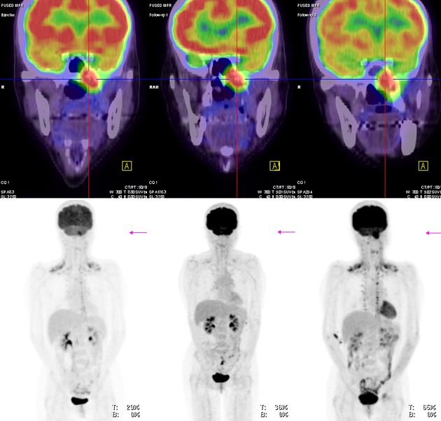
Prominent increased uptake is present at the left side of the base of skull / upper nasopharynx.
Case Discussion
Patient went on to have surgery which confirmed the a cerebral abscess.
Diagnosis of nasopharyngeal carcinoma was established previously on the basis of biopsy (T4N1) 2 years before this presentation. Chemotherapy one year later. Now on Paclitaxel.


 Unable to process the form. Check for errors and try again.