
Presentation
Bilateral lower limb weakness.
Patient Data
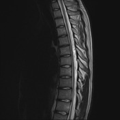


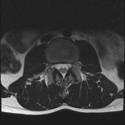

The vertebral bodies have normal alignment, height, and bone marrow signal. Incomplete posterior fusion at L5 and S1.
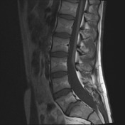
Widening of the bony spinal canal from L3/4 to S1/2 due to dural ectasia. No spinal canal stenosis or cord compression.
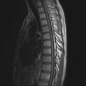
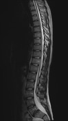
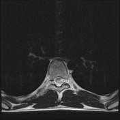
The spinal cord has normal signal and morphology along the thoracic spinal canal, being positioned more anteriorly. The conus medullaris terminates at a lower position, at the level of L3, and is associated with a slightly ticked filum terminale. No signs of associated lipoma or myelomeningocoele.
Intervertebral discs are normal and the intervertebral foramina are capacious.
Conclusion: Lower lumbar dural ectasia and tethered cord.
Case Discussion
Classically, tethered cord on imaging manifests as a low conus medullaris (below L2) and thickened filum terminale (> 2 mm).


 Unable to process the form. Check for errors and try again.