
Presentation
Routine screening mammogram. Asymptomatic.
Patient Data

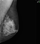
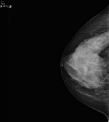
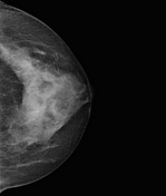

There is dense fibroglandular tissue throughout both breasts. A bilobed circumscribed mass is present in the inferior central right breast.
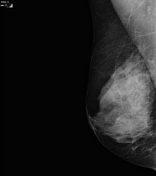
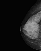

Comparison with the prior right breast mammogram from the previous screening round indicates that there has been interval increase in size of the inferior breast mass.
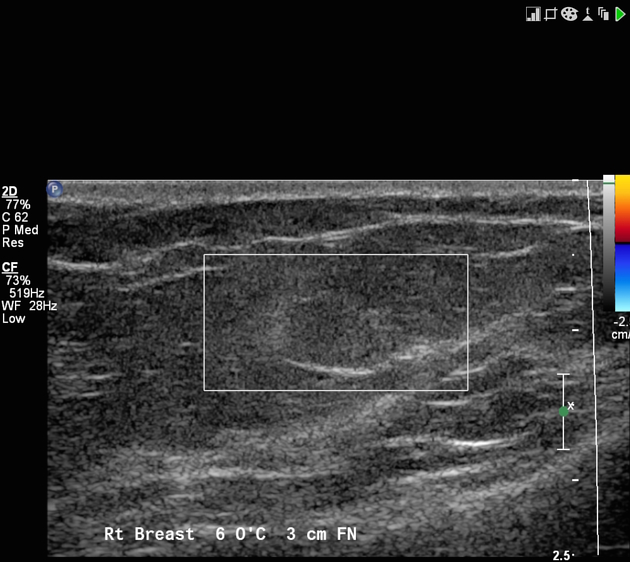
There is a subtle, near iso-echoic circumscribed mass in the right breast 6 o'clock 3 cm from nipple, without internal flow on Doppler assessment.
This corresponds to the mammographic lesion shown above.
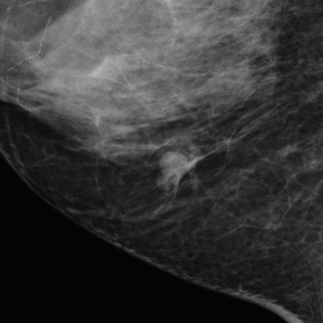
Magnified view better displays the mammographic lesion.
US-guided core biopsy pathology report:
Richly vascularized lesion, 4mm across, circumscribed edges. Variably sized crowded capillary vessels and cavernous vascular spaces are seen, in part with intraluminal RBCs and occasional mononuclear leukocytes. The intervening stroma is minimally edematous and fibrous. There is no solid endothelial proliferation, necrosis or pseudopapillary hyperplasia.
There is no complex infiltrative pattern, cytological atypia or mitotic activity.
Comment:
The sharp circumscription and lack of atypical features favors a benign hemangioma.
The possibility of angiosarcoma should be considered with any vascular lesion of the breast, requiring clinical and radiological correlation.
Case Discussion
The mammographic mass has a circumscribed margin, most suggestive of a cyst. However, it has increased in size since the prior study and there are no other visible circumscribed masses in either breast (i.e. appearance does not fit the "multiple bilateral masses" category 1,2) - hence prompting further assessment.
At ultrasound, it was subtle, near isoechoic and superficial.
Both the mammogram and ultrasound appearances are compatible with a hemangioma, however given the relative infrequency of this diagnosis, other differentials would generally be considered first.


 Unable to process the form. Check for errors and try again.