
Presentation
Renal transplant
Patient Data
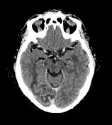

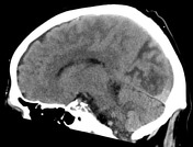
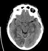


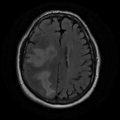
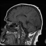
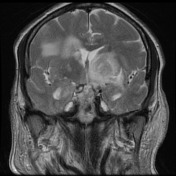
There are multiple ring-enhancing lesions within the brain with the largest situated in the right occipital lobe with surrounding vasogenic edema. There are smaller lesions in the right parietal lobe, right frontal lobe, left basal ganglia and right periventricular location. The remaining lesions measure approximately between 12 and 14 mm in transverse diameter with mild to moderate surrounding vasogenic edema. There is mild effacement of the ventricular system. There is no extra axial fluid collection or any blood in the subarachnoid space. The posterior fossa is normal. There is no cerebellar involvement. The calvarium and skull base are normal. There is no significant paranasal sinus mucoperiosteal thickening.
CONCLUSION:
Multiple intra cerebral focal lesions with ill-defined ring enhancement and associated vasogenic edema. In the appropriate clinical context, these lesions may represent multiple abscesses but equally, metastatic disease cannot be excluded.
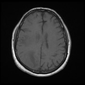

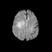
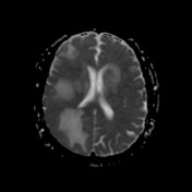
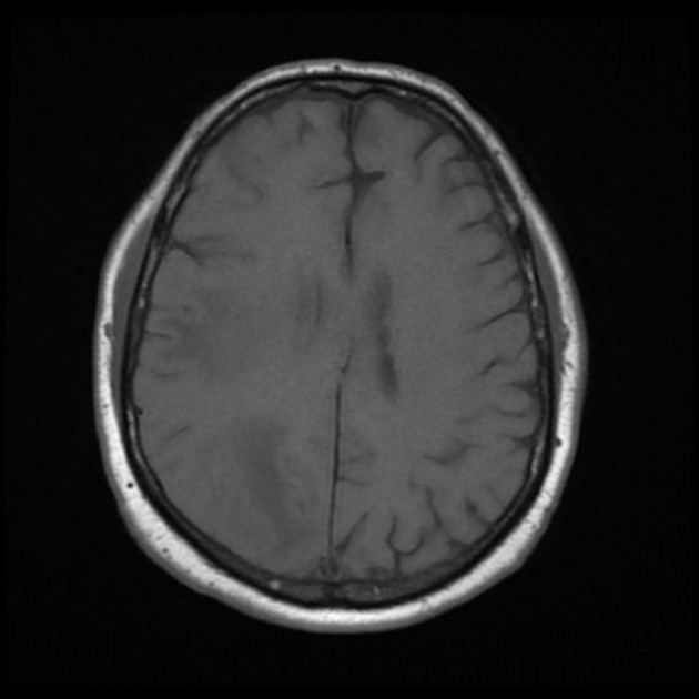
Multiple intracranial masses with pronounced surrounding vasogenic edema are again identified. The masses themselves are isointense or mildly hyperintense to cortex on FLAIR and T2-weighted imaging, in contrast to the surrounding markedly T2 hyperintense vasogenic edema.
The more rounded lesions include: Left lentiform nucleus. Left infralateral thalamus. Left superomedial thalamus. Left posteromedial temporal lobe. Right corona radiata . Right posterior frontal subcortical. Left superior frontal gyrus contains at least two small cortical lesions with subjacent vasogenic edema.
On the CT performed yesterday these rounded lesions were centrally iso to hypodense with peripheral enhancement. The left thalamic lesion demonstrates internal marked ADC reduction of non-enhancing content which raises the possibility of abscess. Linear diffusion restriction related the right corona radiata and right posterior frontal cortical abnormality appears to correspond to locations demonstrating enhancement on CT.
In addition, there is cortical expansion with pronounced subjacent vasogenic edema throughout the right occipital lobe, where thick leptomeningeal enhancement was documented on recent CT. A region of right frontal vasogenic edema and cortical signal abnormality was also associated with leptomeningeal rather than rounded focal enhancement on recent CT. There is no midline shift or uncal herniation. No tonsillar herniation. No hydrocephalus.
CONCLUSION:
Multiple intraparenchymal masses and areas of leptomeningeal abnormality with associated vasogenic edema, corresponding to the areas of thin walled peripherally enhancing masses in leptomeningeal enhancement on recent CT. In the context of immunosuppression, atypical infection (including toxoplasmosis and fungus) is favored over metastasis. The left thalamic lesion and to a lesser extent left lentiform nucleus lesion demonstrates internal ADC reduction corresponding to non-enhancing content, which raises suspicion for cerebral abscess.
Case Discussion
The patient went on to have a biopsy.
Histology
MICROSCOPIC DESCRIPTION: Paraffin sections show fragments of cerebral cortex and white matter. Within the white matter there is aggregation of lymphocytes and plasma cells with foci of necrosis containing acute inflammatory and necrotic debris. Scattered small aggregates of degenerate organisms with features strongly suggestive of tachyzoites of Toxoplasma are noted. The adjacent white matter and cortex is edematous and shows foci of active cerebritis. No evidence of tumor is seen.
FINAL DIAGNOSIS: Cerebral toxoplasmosis.
Serology
Toxoplasma IgG Antibody: DETECTED


 Unable to process the form. Check for errors and try again.