Presentation
Elevated levels of prolactin.
Patient Data
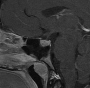
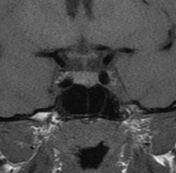


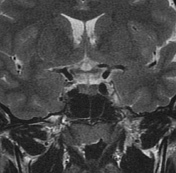
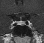

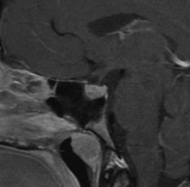
The pituitary gland has normal dimensions and shows a nodular region of delayed enhancement in its anteroinferior aspect on the left, measuring up to 5.5 mm. The infundibulum is centered and has normal thickness. Optic chiasm and suprasellar cistern are unremarkable.
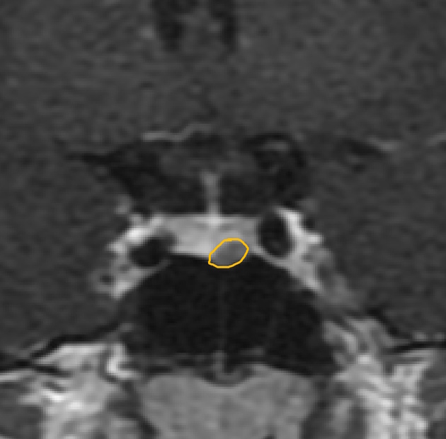
Coronal T1 C+ delineating the nodular area of delayed enhancement in the inferior aspect of the pituitary gland.
Case Discussion
This is a common daily finding for those radiologists reporting pituitary MRIs: a pituitary microadenoma.
It is defined as a nodular area of delayed enhancement within the pituitary gland, measuring less than 10 mm. For this patient, due the elevated prolactin levels, this most likely represents a micro prolactinoma, which is a functioning pituitary adenoma.


 Unable to process the form. Check for errors and try again.