
Presentation
Chronic lower back pain.
Patient Data


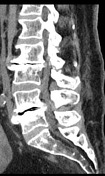
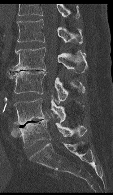
Findings: Five lumbar vertebral bodies with normal height and alignment. There is diffuse osteopenia and degenerative changes, in special within the L2/3 and L4/5 levels, in which prominent intervertebral osteochondrosis are noted: irregularities and subchondral sclerosis involving the endplates, marginal osteophytes, and disc degeneration with gas.
L1/2: Disc has normal height and density. Mild posterior disc bulging indenting the dural sac anteriorly. No foraminal impingement.
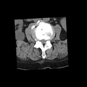
L2/3: Osteochondrosis as previously described. Disc bulging associated with osteophytes compressing the dural sac anteriorly and moderately stenosing the right intervertebral foramen, in which there is impingement of the right L2 exiting root.
L3/4: Disc has normal height and density. Disc bulging compressing the dural sac anteriorly and stenosing the intervertebral foraminal bases, touching the left L3 exiting root.
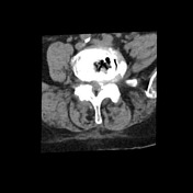
L4/5: Osteochondrosis as previously described. Disc bulging associated with osteophytes compressing and stenosing the dural sac anteriorly and severely stenosing the left intervertebral foramen, in which there is impingement of the left L4 exiting root.
L5/S1: Disc has normal height and density. Intervertebral foramina are capacious.
Conclusion: L2/3 and L4/5 osteochondrosis with disc-osteophyte complexes stenosing the spinal canal focally and promoting impingement of the right L2 and left L4 exiting nerve roots. Less prominent degenerative changes in the other levels, as described above.
Case Discussion
This case illustrates a severe L2/3 and L4/5 intervertebral osteochondrosis with disc-osteophyte complexes stenosing the spinal canal locally and promoting impingement of the right L2 and left L4 exiting nerve roots. Less prominent degenerative changes in the other levels, as described above.


 Unable to process the form. Check for errors and try again.