
Presentation
Right eye epiphora for six months
Patient Data
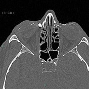

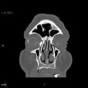
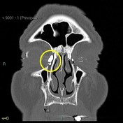
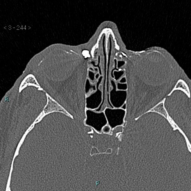
Post saccular narrowing from prior iatrogenic tear post-dacryocystitis.
The left nasolacrimal duct is conveying the contrast medium into the nasal cavity, and then the nasopharynx.
Case Discussion
The nasolacrimal duct normally drains tears from the eye down into the nose.
The opening of the duct is in the medial canthus of the eye.
One of the most frequent problems of the lacrimal pathways is stenosis and blockage of the tear flow.
Complications of post-saccular stenosis are infections with abscess formation that require drainage.
The stenosis may be congenital, in which case they may present in neonates.
The acquired strictures typically happen in adults with a known history of ipsilateral nasal pathology due to prior trauma or an obstructing mass lesion.


 Unable to process the form. Check for errors and try again.