
Presentation
Microscopic hematuria. The patient recently had a CT urogram in which there was nonfilling of the right distal ureter, so a retrograde pyelogram was ordered.
Patient Data
Age: 65 years
Gender: Male
From the case:
Ureteral pseudodiverticulosis

Download
Info
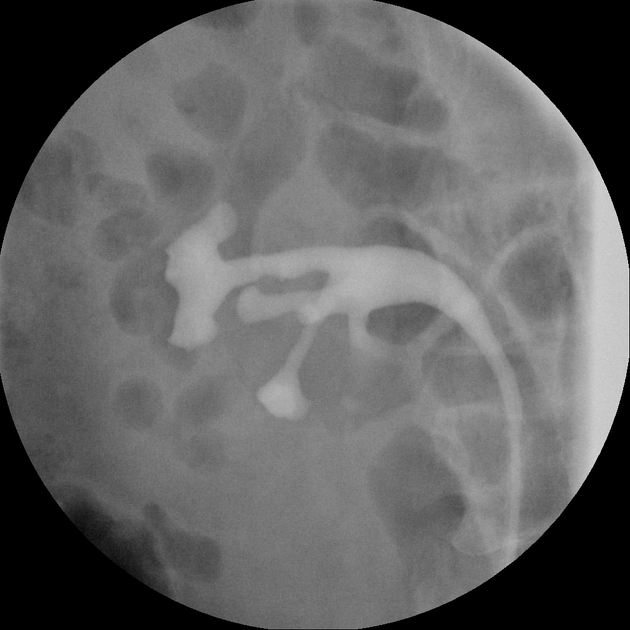
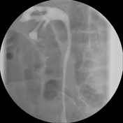
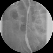
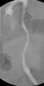
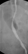
Images from a retrograde pyelogram: In the mid-ureter, there are tiny (1-2 mm) outpouchings of contrast, compatible with ureteral pseudodiverticulosis.
From the case:
Ureteral pseudodiverticulosis


Download
Info
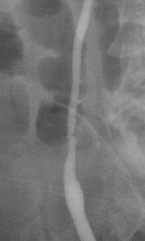
Arrows point to the multiple tiny pseudodiverticula in the mid-ureter.
Case Discussion
Ureteral pseudodiverticulosis is an uncommon process that has been associated with the development of urothelial (transitional) cell carcinoma.
It can be seen on a CT urogram (CTU) but is much easier to detect on IVU or a retrograde pyelogram. In this case, the pseudodiverticula was not visible on the CTU.


 Unable to process the form. Check for errors and try again.