
Presentation
Right suprarenal cystic lesion discovered on sonography.
Patient Data


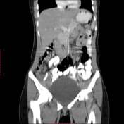

A well-defined right adrenal cystic lesion is seen measuring about 6 cm in diameter with internal fluid attenuation reading about 25 HU. A mural based isodense nodule is seen at the infero-medial wall measuring about 2 cm.
Case Discussion
A right adrenal cystic lesion with a mural based isodense nodule suggestive of a complex adrenal cyst.
Surgical excision was performed grossly showing a right adrenal cystic mass with a wall thickness 0.2-0.4 cm, smooth surface with a well defined soft tissue like brownish material within measuring 2.5 x 1.5 cm. Microscopic appearance showed a fibrotic wall with no epithelial or endothelial lining. The cyst contains only blood with no malignancy detected.


 Unable to process the form. Check for errors and try again.