
Presentation
Unwitnessed fall on the ward.
Patient Data
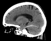

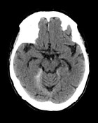

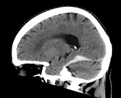
There is an acute subdural hematoma observed along the superior portion of the tentorium on the right, measuring up to 3.2 mm in depth.
No intra-axial collection, mass or focal abnormality identified. Ventricles and remainder basal cisterns are unremarkable. Patchy white matter low attenuation is non-specific, but most likely represents sequelae of chronic small vessel ischemic change, and is of an amount commonly seen in this age group.

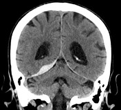
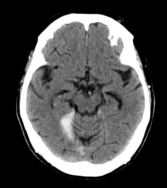
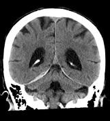
There has been progression of the right subdural hematoma tracking along the right tentorium now measuring 9 mm at its greatest depth ( previously 3.2 mm ) and extends to the level of the right anterior clinoid process.
No new intra or extra-axial hemorrhage is identified. The ventricles and basal cisterns are unremarkable. Grey-white matter differentiation is preserved with no midline shift or mass effect. No abnormality of the posterior fossa is identified. Visualized paranasal sinuses and mastoid air cells are clear.
Case Discussion
This case illustrates a typical incident that can happen with inpatients, especially among the elderly, and that is usually targeted by numerous institutional prevention campaigns. Unfortunately, even with all the prevention measures, it is not uncommon to receive requests for CT scans similar to this case.
First CT scan demonstrated a right-sided subdural tentorial hematoma, hematomas like this one may sometimes be really thin and just identified by a subtle hyperattention of the tentorium. The follow-up scan performed one day later has demonstrated progression over the hematoma, but no further mass effect or other complications.


 Unable to process the form. Check for errors and try again.