
Presentation
Left medial periorbital painless subcutaneous swelling since birth.
Patient Data


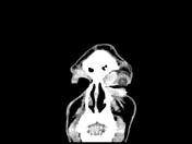
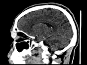
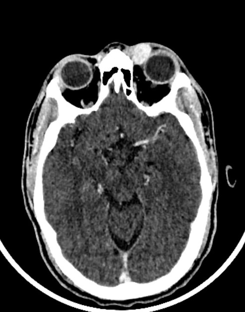
Oval shaped left intra-orbital extra-conal lesion currently measuring 12 x 16 x 17 mm with well-defined outlines. It is located along the superomedial aspect of the left orbit just inferior to the left superior oblique muscle, insinuating itself between the superior-medial bony orbital rim and the globe. The latter is mildly displaced laterally. The lesion showed avidly enhancing matrix. No related significant bony changes. The left orbital structures are otherwise, normal.
Case Discussion
The described CT features are suggestive of the left orbital extra-conal lesion, mostly representing capillary hemangioma of the orbit.
Capillary hemangioma of the orbit is different from orbital cavernous hemangioma. It is usually present since birth anterior to the globe with imaging characteristics of avid homogenous enhancement in contrast-enhanced CT.


 Unable to process the form. Check for errors and try again.