
Presentation
Shortness of breath and chest discomfort during exertion.
Patient Data
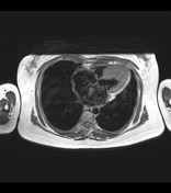

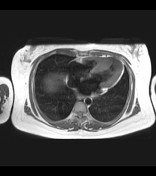

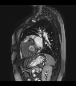
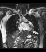
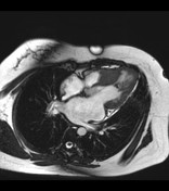

There is small left ventricular cavity size with thickened walls, most pronounced at the septal region. Findings are consistent with asymmetric hypertrophic cardiomyopathy. Cine images show near complete, systolic, mid ventricular cavity obliteration, with intermittent systolic anterior motion of the chord and anterior mitral leaflet suggestive of dynamic outflow obstruction.
Case Discussion
This is a cardiac MRI of a patient with asymmetric hypertrophic cardiomyopathy. There is associated systolic anterior motion (SAM) of the anterior mitral leaflet, which contribute to left ventricular outflow tract (LVOT) obstruction. Hypertrophic cardiomyopathy is a heterogeneous group of diseases related to sarcomere gene mutations and affects one of every 500 adults. It is the most common cause of sudden death in young athletes. Asymmetric hypertrophic cardiomyopathy is the most common morphologic variant of this disease. This MRI was requested for evaluation prior to contemplated myomectomy.


 Unable to process the form. Check for errors and try again.