
Presentation
Non-specific headaches
Patient Data
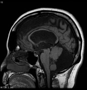

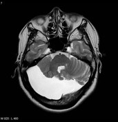
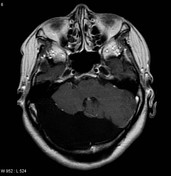


MRI through the posterior fossa demonstrates a large right-sided extra-axial CSF intensity mass lesion. It follows CSF on all sequences, including FLAIR and DWI/ADC. There is significant mass effect on the adjacent cerebellar tissue and remodeling and expansion of the adjacent skull is evident.
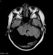
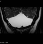
High resolution T2 images (FIESTA) demonstrates that this lesion is bounded by a very thin membrane, best seen bulging across the midline towards the left.
Sagittal images demonstrate upward bowing and thinning of the corpus callosum suggesting hydrocephalus.
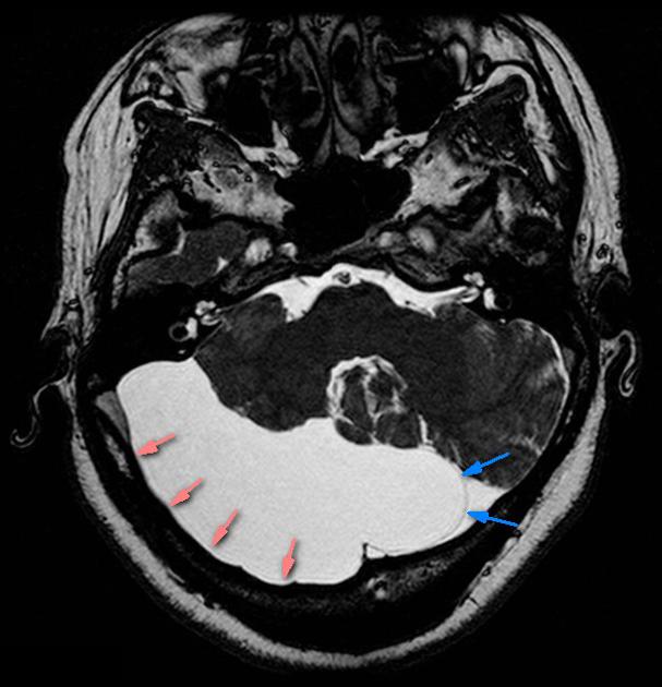
The membrane of the arachnoid cyst can be seen bulging to the left of the midline (blue arrows).
Remodeling of the skull is also present (pink arrows).
Case Discussion
This case illustrates many of the features of arachnoid cysts, although actual visualization of the membrane as clearly as this is not always achieved.


 Unable to process the form. Check for errors and try again.