
Presentation
Slowly enlarging, non-painful left neck mass. No other palpable mass lesions.
Patient Data
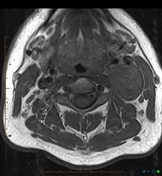
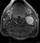

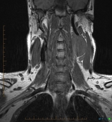
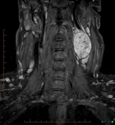

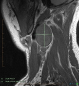
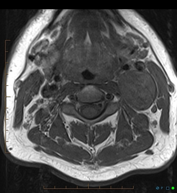
A large, well-defined, mass with diffuse enhancement sitting posterior to and displacing the common carotid artery and carotid bifurcation, as well as the internal jugular vein, anteriorly.
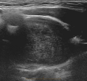
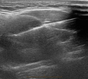

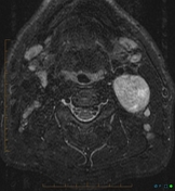
Rounded well-defined, solid mass in the left side of the neck with biopsy needle in situ.
Case Discussion
Histology
Pathology result shows a tumor of bland spindle cells devoid of cytological atypia but with nuclear pallisading. These cells stain with S100. Appearances are typical of schwannoma.
Mass lesions intimately related to the carotid bifurcation include lymph nodes, paragangliomas and schwannomas.
Given that this is an isolated lesion, a lymph node seems unlikely but possible.
Paragangliomas are vascular and contain flow voids (salt and pepper appearance).
Schwannomas tend to be a well-defined mass as in this case. It is likely arising from the vagus nerve given its location posterior to the carotid bifurcation (the vagus nerve runs along the posterior aspect of the carotid sheath).


 Unable to process the form. Check for errors and try again.