
Herniation of the optic chiasm and third ventricle into a partial empty sella
Presentation
Known history of pituitary prolactinoma a few years ago. Treated conservatively with dopamine agonists. Lost clinical follow-up, presenting now with visual impairment.
Patient Data
MRI Pituitary gland
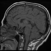

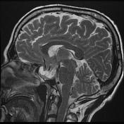
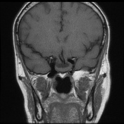
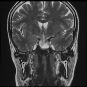
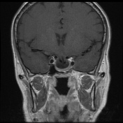
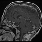
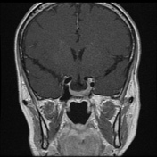
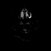


There is a prominent enlarged sella, which is partially empty, and has an associated downward herniation of the floor of the third ventricle, optic chiasm and proximal segments of the anterior cerebral arteries within. The pituitary gland shows to be relatively enlarged and displaced peripherally against the sellar walls.
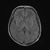
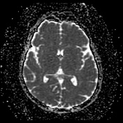
The remainder of the brain is unremarkable.
Case Discussion
Herniations into the sella have been described associated either with primary or secondary empty sella. Such as in this case, visual disturbances have been well documented associated with secondary empty sella (medical/surgical therapy of a sellar tumor like marcoadenoma).
Unfortunately, previous imaging were not available to make a clear correlation with the tumor (documented macroadenoma).


 Unable to process the form. Check for errors and try again.